Project Description
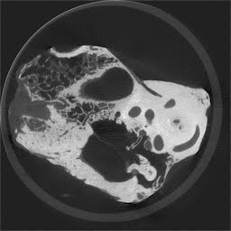
The middle-ear anatomy is integrally linked to both its normal function and its response to disease processes. To improve our understanding of middle-ear anatomy, we require an intuitive and effective method to examine its morphology in a non-destructive manner. The advent of volumetric CT has made it possible to image features of interest relatively quickly and without the need for physical sectioning. While images acquired using standard clinical CT scanners lack the spatial resolution needed to fully capture the intricacy of middle-ear anatomy, micro-computed tomography (micro-CT) imaging provides high-resolution anatomical data that accurately represent finely detailed structures such as the ossicles.

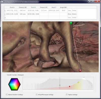
We present a software and hardware suite with the objective of overcoming some of the limitations of existing methods. We have developed a system with interactivity and ease of use in mind that allows clinicians and scientists to explore and analyze micro-CT scans of the middle ear. By combining state-of- the-art software and hardware technologies such as direct volume rendering, haptic feedback, virtual surgical microscope navigation, and a user-friendly interface, we seek to create an interactive morphometric workstation that allows the user to quickly and intuitively explore micro-CT data and perform quantitative measurements directly on the anatomical structures of interest.

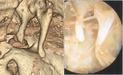
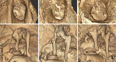
This software represents an effective method to reconstruct and explore otological micro-CT datasets for education and research, and to yield greater understanding of relevant middle-ear functional anatomy. Examination of micro-CT datasets of cadaver temporal-bone specimens show that the reconstructed images accurately represent the fine features of middle-ear anatomy.

We used our software to examine three different micro-CT scans of human temporal bones. Considerable detail can be seen of functionally relevant anatomy. We found that the use of the haptic interface device to control a virtual tool allowed the user to quickly and accurately place measurement markers on specific structures and anatomical features.

The ability to load and visualize micro-CT middle-ear datasets makes this a powerful educational tool for learning the intricacies of middle-ear anatomy. Such fine structures as the ossicles and their ligamentous attachments are not adequately rendered from clinical CT scans, and normally not appreciated until the trainee first encounters them through the operating microscope on an actual patient. Our visualization suite is an interactive anatomy lesson for residents and medical students. By exploring the middle-ear space at high resolution, the trainee can quickly grasp the 3D relationships between its structures and achieve a sense of their shape and scale.
We expect that, with additional validation, these technologies will enable further insights into otological function and disorders. The technologies may contribute to clinical care through improving the understanding of salient anatomy, improving diagnosis and pre-operative planning, and aiding in the optimal design and fitting of ossicular reconstructive materials or implantable hearing devices. Similarly, through applying these technologies to biomechanical modeling, they may also contribute to our understanding of the basic functioning of the hearing mechanisms.
Related Publications
Lee, D. H. et al. Reconstruction and exploration of virtual middle-ear models derived from micro-CT datasets. Hearing Research (2010).
Project Staff
Status
Active since 2002.
Funding Sources
This project was funded in part by NIH Grant 5R01LM010673-02
and in part by the Veterans Administration.