Project Description
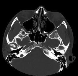
>Endoscopic sinus surgery (ESS) is a technically challenging endeavor that could benefit from a rehearsal environment. Important structures such as the orbit and its contents, optic nerve, carotid artery, cranial vault, and nasolacrimal system are at risk for injury. A thorough understanding of the anatomic variability encountered during surgery is critical in achieving successful outcomes and decreasing complications.
 |
 |
Angles of surgical approach and regions of limited access can be difficult to envision given a series of standard two-dimensional slices. Currently there is no intuitive method for interacting with preoperative imaging data. New technology to allow surgeons to view, manipulate, and even feel a patient’s anatomy based on preoperative imaging could provide considerable improvement over traditional planning methods.
 |
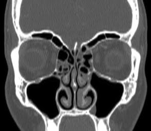 |
A 3-dimensional virtual surgical environment may allow for enhanced pre-operative planning and rehearsal, with the goal of improving patient outcomes, decreasing complication rates, and enhancing technical skills. The purpose of this study is to report on the design, features, and initial experience with the Stanford Virtual Surgical Environment (VSE) for rhinologic procedures. The VSE allows a surgeon to interact with patient-specific, 3-dimensional reconstructions of sinus CT datasets using a modified haptic (touch feedback) interface device driving a virtual endoscope.
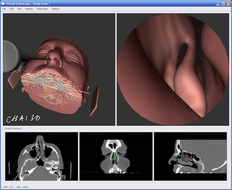
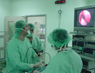
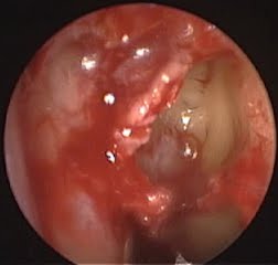
Initial evaluation of the VSE was then performed using the CT datasets from 2 patients who underwent endoscopic sinus surgery. The patients’ surgical procedures were recorded to enable comparisons between the virtual endoscopic views and actual intraoperative endoscopic views. Three experienced surgeons performed endoscopy on the virtual representation of the patient to produce rendered images to match the intraoperative views. Using the intra-operative video as a reference, the virtual endoscope was navigated through the nasal cavity to the site of the procedure. Images of the virtual procedure were captured during this process to facilitate comparison. Anatomical correlations, key structural features, and other similarities between the intra-operative images and the simulated procedure were noted.

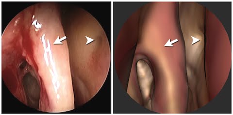
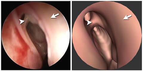
The VSE allowed for high fidelity anatomic correlation between virtual and intraoperative findings based on the surgeons’ subjective impression, and the comparison of virtual and intraoperative images. The graphics rendering frame rate approximates that of smooth endoscopic video. A key element in the design of the Stanford VSE is the capability to directly load pre-operative CT DICOM image series to reconstruct a “virtual patient” in 3D. Our VSE offers the surgeon the ability to interact with this virtual patient through a simulated endoscope similar to that used in ESS. Patient-specific surgical simulation offers a new frontier in patient care and medical education.
Related Publications
Parikh, S. S. et al. Integration of patient-specific paranasal sinus CT data into a virtual surgical environment. American Journal of Rhinology (2009).
Project Staff
Status
Active since 2002.
Funding Sources
This project was funded in part by NIH Grant 5R01LM010673-02
and in part by the Veterans Administration.